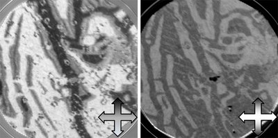
Figure 1 below shows two dichroic PEEM images (field of view 20 um), using the XMCD and XMLD effects as magnetic contrast mechanisms. On the left, using XMCD at the Fe-L3 absorption edge, the dominating four-fold anisotropy of the Fe3O4 crystal can be appreciated. The intensity grey scale corresponds to the magnetization orientation relative to the incoming X-ray beam as indicated by the inset arrow. The right image was taken using linear horizontal polarized X-rays at two different energies at the Fe-L2 absorption edge. For the same magnetization pattern as in the left image, the different angle dependence of the XMLD effect (inset arrow) results in the same color for antiparallel domains.

Fig. 1: XMCD (left) and XMLD (right) magnetic microscopy images of the same zone in a Fe3O4 single crystal (sample courtesy of A.K. Schmid and Juan de la Figuera). The PEEM images (field of view 20 um) were taken at the CIRCE beamline using photons with either circular or linear horizontal polarization tuned to the Fe-L3 (XMCD) and Fe-L2 (XMLD) absorption edges. The intensity grey scale represents the orientation of the magnetization as indicated by the inset arrows.
Matteo Cantoni, Lorenzo Baldrati, Christian Rinaldi and Riccardo Bertacco (Figure 2) from the Politecnico de Milano have used the CIRCE-PEEM endstation to investigate magneto-electric devices based on a single, 1.1 nm thin, CoFeB layer with perpendicular magnetic anisotropy (PMA) on ferroelectric BaTiO3.

Fig. 2: Lorenzo Baldrati (left) and Matteo Cantoni (third from left) with ALBA staff.
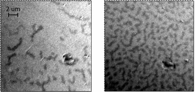
Figure 3 below shows the magnetic configuration of the same part of a sample in different remanent states, exhibiting typical worm-like domains of PMA materials. A new sample holder with in-built electromagnet was used for in-situ magnetization reversal of the samples, allowing us to take images in different remanent states and with magnetic field applied. Due to the near grazing x-ray beam incidence in the PEEM, the realization of out-of –plane magnetic contrast is more challenging than in-plane contrast. Future studies will address the influence of the ferroelectric BaTiO3 on the magnetic switching of the top CoFeB structure.

Fig. 3: XMCD-PEEM image taken at the Fe-L3 absorption edge of a CoFeB layer in two different remanent states. The grey scale contrast corresponds to the direction of the perpendicular magnetization (up or down), and arises from the different projections on the photon beam illuminating the sample under 16° grazing incidence. Different remanent states were realized by applying magnetic fields with an electromagnet integrated in the sample holder.


