

FAXTOR is the new micro-tomography beamline dedicated to Fast X-ray Tomography & Radiography currently under development at ALBA. FaXToR will be a versatile beamline aiming at performing quasi-real-time 3D computed tomography. In particular, in addition to the traditional 3D computed tomography, FaXToR will represent a new tool for the user community to study the dynamics of certain processes at the micrometric scale using X-rays. In order to achieve this goal, the beamline requires high fluxes and excellent beam quality. The possible fields of applications are multifold and include material science, biology, paleontology, earth science, cultural heritage and industrial application.
Technique
Propagation-based phase-contrast and absorption-based imaging techniques will be made available to the users at the beamline, this will allow exploiting the absorption and refraction properties of a sample at the micrometric scale. Grating-based method will provide simultaneously three signals: the absorption contrast, the differential phase contrast and the dark field. Radiography and fast/slow computed tomography will be the available acquisition protocols.
Energy parameters
The beamline is foreseen operating at a range of energies, from 8 keV to 70 keV, in both monochromatic (8-50 keV) and filtered white beam modes (30-70 keV).
STATUS
FAXTOR is currently under construction and commissioning. The multi-pole wiggler and the front end have been successfully installed and commissioned (Figure 1). The optical and experimental hutches have been installed (Figure 2). The control room which will host the workstations for th beamline control and the data acquisition and processing, foreseen a dedicated area for the sample preparation. The optics elements are under manufacturing and commissioning, while the end station is currently under production.
The first user operation is expected by the beginning of 2024.
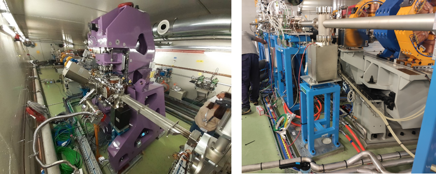
Figure 1: (left) ID installed in the tunnel and commissioned. (Right) FE under installation and commissioning.

Figure 2: (left) Optic Hutch. (Right) Experimental Hutch
TECHNICAL SPECIFICATIONS
| Temporal resolution | 20 Hz Max Tomography |
| Spatial resolution (Pixel size) | 0.5-10µm |
| Energy range | 10 keV – 50 keV (mono) / 30-70 keV (filtered whitebeam) |
| Monochromator | Double multilayer |
| Whitebeam option | Available at the end-station |
| X-ray source | Multipole wiggler |
| Imaging techniques |
Propagation-based imaging, absorption-based imaging. Grating-based imaging. Radiography and Computed Tomography modality. |
OPTICS
Beamline layout
The main optical element of the beamline is represented by a double multilayer monochromator (DMM). Several other optical components like CVD diamond Windows, filters units, slits, beamline diagnostic elements, shutters will complete FAXTOR’s optical layout.
The beamline is divided into two parts, an optical hutch, hosting the DMM and the other optical elements, and the experimental hutch, in which the experiments will be carried out. The sample stage, a 6-axis, will be located at 36 m from the source.
Source
FAXTOR will be feed by a 5 poles in-vacuum multipole wiggler. The peak magnetic field expected is 2.5 T. The ID parameters have been optimized in order to keep a reasonable source size (horizontally) while delivering high fluxes.
Monochromator
The monochromator will be a double multilayer monochromator, with an operational energy range between 8 keV and 50 keV. Multiple multilayer stripes (W/B4C and Ru/C) are foreseen for an optimization of its performances. Thanks to the use of long crystals (500 mm lenght) the beam losses in the vertical dimensions due to the small grazing angles are minimized. The expected energy bandwidth will vary between 2% and 4%, according to the selected stripe.
Beam size
The maximum beam size expected at the entrance of the sample is of 35 mm horizontally, while vertically it will range between 5 and 12 mm, according to the energy selected/energy spectrum.
Microscope
FAXTOR will make use of microscopes based on the indirect detection of X-rays, that coupled with fast acquisition cameras (CMOS) will represent the beamline detection system. Multiple magnifications will be made available allowing performing acquisitions at different spatial resolutions within a range of 0.5 – 10 µm (image pixel size) and a field of few varying from few mm to few cm. It will be possible to optimize the sample-to-detector distance according to the experimental needs within a range between ~0 and 4 m.
Sample
Sample sizes can vary from a few mm to a few cm.
The tomographic stage will be compatible with custom sample environments allowing performing in-situ/in-operando measurements.
On top of the detection table, an auxiliary platform will be accommodated to allow complex in-situ devices.


