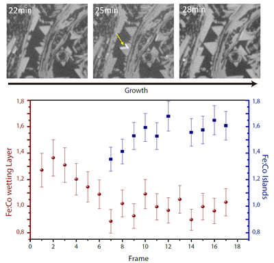
Figure: XAS PEEM images at different stages of growth of a Fe-Co oxide on Ru. The time elapsed is indicated in the upper left label on each image. As can be seen in the images, the islands grow in size, often with triangular shape. The graph shows the composition of the wetting layer and the islands during growth.
Cerdanyola del Vallès, 25th March 2020. Researchers from the Universidad Complutense de Madrid, the Institute of Ceramics and Glass (ICV-CSIC), the SURFMOSS group at the Instituto de Química Física Rocasolano (IQFR-CSIC) and the ALBA Synchrotron have studied the dynamic changes in the distribution of cobalt (Co) and iron (Fe) atoms during the growth of mixed cobalt-iron oxides on a metallic substrate.
The preparation method, oxygen-assisted molecular beam epitaxy, has been shown to produce thin films and nanostructures of transition metal oxides with much higher structural quality than those obtained by other methods. In turn, this results in magnetic properties closer to the ideal ones, making the films very promising for applications for example in spintronics.
The initial composition is a mixed Fe-Co (II) oxide wetting layer reflecting the ratio of the deposited materials. However, as subsequent growth of three dimensional spinel islands nucleating on this wetting layer takes place, the composition of the oxide in the wetting layer changes as iron is transferred into the spinel islands. The composition of the islands themselves also evolves during growth.
During the growth by high temperature oxygen assisted molecular beam epitaxy, fast X-ray PhotoEmission Electron Microscopy sequences were recorded scanning the photon energy through the Fe and Co absorption edges. Thus spatial variations of the composition during the growth were resolved in real time.
These results have been obtained at the CIRCE beamline of ALBA, by low-energy electron microscopy (LEEM) and photoemission electron microscopy (PEEM) techniques.
This is the first time that growth at such high temperatures (1.000 ºC) has been followed in real space and real time with synchrotron radiation, providing valuable dynamic chemical information that would have otherwise been impossible to detect.


