A research group from the Weizmann Institute of Science from Israel is investigating the cholesterol crystals' formation. There is a link between cholesterol crystal accumulation, cell debris (plaque) presence inside the blood vessels and the patients' survival. Scientists have been enriching with cholesterol macrophages, immune system cells that are involved in the formation of atheroma plaques. The deposition of excessive cholesterol in these plaques, which block the bloodstream, is a prominent feature of atherosclerosis, a major precursor of many cardiovascular diseases.
These macrophages with cholesterol inside them are the cause of the inflammation that occurs before the atheroma plaque. They are attached in the blood vessel walls and, while they are still alive, produce cholesterol crystals that are excreted from the cells. Finally, macrophages end up dying and releasing the rest of cholesterol crystals they have inside. However, the crystals' formation mechanism is still not well-understood. This information will help to discover how to inhibit the process and develop more effective treatments for atherosclerosis.
To carry out the experiment, scientists have brought their samples to the ALBA Synchrotron. They have been using the soft X-ray microscopy beamline, MISTRAL, and tomographies (3D images) of macrophages with the cholesterol crystals have been obtained. Researchers had already seen in vitro that cholesterol crystals (three-dimensional) can be nucleated from two-dimensional crystalline domains, located in cell membranes. Verifying this hypothesis in a biological system and visualizing macrophages secreting the crystals was their main objective.


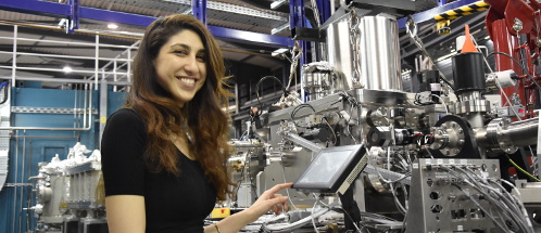
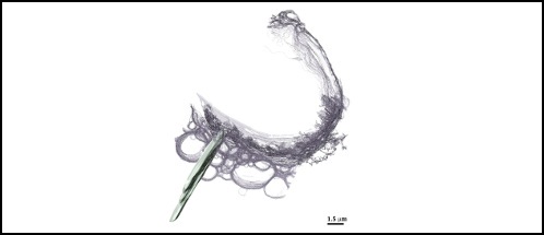
Left: Neta Varsano, one of the group researchers, in front of the MISTRAL beamline. Right: tomography of a cell (plasma membrane in purple) that generated large cholesterol crystal (segmented in green). Image obtained in MISTRAL.
MISTRAL beamline equipment plays a key role in this type of experiments as it enables to obtain tomographies of whole cells. In other words, the MISTRAL microscope is non-invasive: there is no need to section the cell in thin layers, so that the whole sample can be observed close to physiological conditions. There are only three more beamlines like MISTRAL all over the world (UK, Germany and USA).


